Patient Guides & Medical Insights

Comprehensive surgical guides and educational resources by Dr. Gopal Sharma, MCh, MRCS — Uro-Oncology Specialist, Manipal Hospital Dwarka, Delhi.

Patient Guide: Robotic Partial Nephrectomy (RAPN)
KIDNEY CANCER • ROBOTIC SURGERY

Patient Guide: Robotic Partial Nephrectomy (RAPN)

By Dr. Gopal Sharma, MCh MRCS Manipal Hospital, Dwarka

Saving the Kidney: Robotic Partial Nephrectomy

Saving the Kidney: Procedure & Risks

Robotic Partial Nephrectomy is a minimally invasive surgery to remove a renal tumor (mass) while preserving the rest of the kidney.

It is the preferred treatment for small-to-medium kidney tumors because saving kidney tissue lowers your risk of developing chronic kidney disease or needing dialysis in the future.

RAPN

Part 1: The Surgical Steps (What Happens Inside)

Part 1: The Surgical Steps (What Happens Inside)

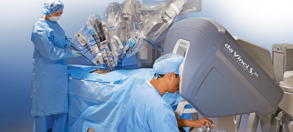
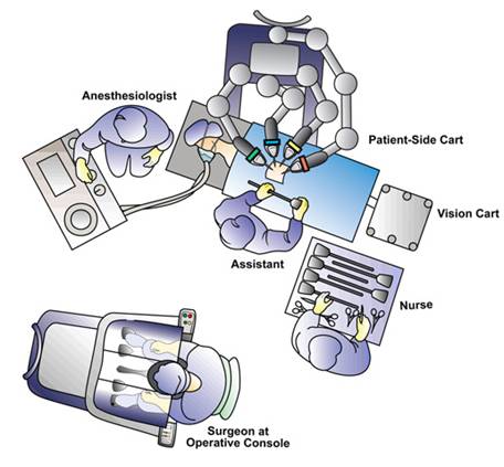
The surgery is performed using the da Vinci Surgical System and typically takes 2 to 4 hours.

Step 1: Positioning & Access

You will be placed on your side (flank position) to give the surgeon access to the kidney.

4–5 small "keyhole" incisions (8–12mm) are made on the abdomen to insert the robotic camera and instruments.

RAPN

Step 2: Exposing the Kidney

The kidney sits behind the intestines. The surgeon carefully moves the colon (bowel) aside to reveal the kidney.

Hilar Dissection: The surgeon locates the main renal artery and vein. This is a critical step because we must temporarily stop blood flow to the kidney to cut out the tumor safely.

Step 3: Defining the Tumor (Ultrasound)

A specialized robotic ultrasound probe is placed directly on the kidney.

This helps the surgeon "see" inside the organ to mark exactly where the tumor ends and healthy tissue begins.

Step 4: Clamping (Ischemia)

Small clamps are placed on the kidney artery to stop blood flow.

"Warm Ischemia Time": This is the clock ticking while the kidney has no blood flow. The robot’s speed and precision allow us to keep this time short (usually under 20–25 minutes) to prevent kidney damage.

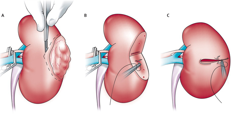
Step 5: Tumor Excision

Using robotic scissors, the tumor is cut out with a thin rim of healthy tissue (margin) to ensure no cancer cells are left behind.

Step 6: Reconstruction (Renorrhaphy)

This is the most complex part. The surgeon must sew the kidney back together in two layers:

Deep Layer: To close the urinary collecting system (preventing urine leaks) and stop deep bleeding.

Outer Layer: To pull the kidney edges together (like closing a clam).

Once sewn, the clamps are removed, and blood flow is restored to the kidney.

RAPN

Part 2: Potential Complications & Risks

Partial nephrectomy is a complex surgery with specific risks related to cutting into a highly vascular organ.

A. Specific Risks of Partial Nephrectomy

1. Bleeding & Transfusion

The kidney filters 20% of the body's blood. While robotic tools minimize blood loss, there is a risk of bleeding during or after surgery.

Risk: Occasionally (<5%), a blood transfusion is needed.

2. Conversion to Radical Nephrectomy (Total Removal)

The Risk: Sometimes, once the surgeon sees the tumor, it may be too close to major blood vessels, too deep, or bleeding too heavily to be saved safely.

The Outcome: For your safety, the surgeon may make the decision during surgery to remove the entire kidney (Radical Nephrectomy) to ensure the cancer is gone and bleeding is controlled. This happens in less than 5% of cases.

3. Urine Leak (Urinoma)

Because the tumor often sits near the urine collection system, cutting it out can create a leak.

Outcome: We usually leave a small drain tube in your side after surgery. If a leak occurs, it usually heals on its own with the drain in place. Occasionally, a ureteral stent (a thin internal tube) must be placed to help it heal.

B. General Surgical Risks

Infection: At the incision sites or urinary tract.

Hernia: A bulge at the incision site (rare with small robotic cuts).

Ileus: Temporary slowing of the bowels, causing bloating or constipation.

Part 3: Recovery & Aftercare

In the Hospital (1–3 Days)

Drain: You will likely wake up with a small drain tube coming out of your side. This monitors for internal bleeding or urine leaks. It is usually removed before you go home.

Catheter: A urinary catheter will be in place overnight and typically removed the next morning.

Pain: Most patients manage well with minimal pain medication due to the small incisions.

At Home

Activity: No heavy lifting (over 10 lbs) for 4–6 weeks. This is critical to prevent the internal kidney stitches from popping open.

Work: Desk jobs can be resumed in 2–3 weeks.

Follow-up: You will need regular CT scans or MRIs for a few years to ensure the tumor does not return

Dr. Gopal Sharma is a skilled robotic surgeon providing one of the most dedicated and specialised treatments for Kidney cancers. He is recognised as one of the top robotic surgeons in Delhi for kidney cancer. Individualised treatment approach with a dedicated surgical team ensures smooth recovery through your difficult times.
Patient Guide: Robotic Radical Cystectomy (RARC)
BLADDER CANCER • ROBOTIC SURGERY

Patient Guide: Robotic Radical Cystectomy (RARC)

By Dr. Gopal Sharma, MCh MRCS Manipal Hospital, Dwarka

Bladder removal: Robotic radical cystectomy

Robotic Radical Cystectomy with Ileal Conduit

This is a minimally invasive procedure to remove the urinary bladder (and nearby lymph nodes/organs) to treat bladder cancer.

Because the bladder is removed, the surgeon creates a new way for urine to leave your body using a small piece of your intestine, called an Ileal Conduit.

Using the da Vinci Surgical System, the surgeon controls robotic arms to perform the operation through small "keyhole" incisions with high precision.

ROBOTIC PORTS

The surgery typically takes 4 to 6 hours and is performed under general anesthesia (you are asleep).

Step 1: Access & Preparation

Inflation: The abdomen is gently inflated with gas (carbon dioxide) to create space for the surgeon to see and work.

Port Placement: Instead of one large incision, 5–6 small incisions (less than 1 inch) are made. The robotic camera and instruments are inserted through these "ports."

RARC

The Surgical Procedure (Step-by-Step)

RARC

Step 2: Removal (The Cystectomy)

Dissection: The surgeon carefully separates the bladder from surrounding structures (intestines, blood vessels).

For Men: The prostate and seminal vesicles are usually removed.

For Women: The uterus, fallopian tubes, ovaries, and part of the vagina may be removed.

Lymph Node Removal: The surgeon removes lymph nodes from the pelvic area. These are sent to the lab to check if cancer has spread.

Extraction: The bladder and organs are placed in a secure bag and removed through one slightly larger incision (usually near the belly button).

RARC

Step 3: Reconstruction (The Ileal Conduit)

Isolation: The surgeon selects a short segment (about 6 inches) of the small intestine (ileum). This piece is disconnected from the rest of the bowel, which is then reconnected so digestion can continue normally.

Connection: The tubes from your kidneys (ureters) are sewn into one end of this isolated intestine segment.

Stoma Creation: The other end of the intestine segment is brought out through an opening in the abdominal wall to form a stoma.

Stents: Small plastic tubes (stents) are placed inside the ureters temporarily to help the connection heal.

Complication Description
Ileus (Slow Bowel) The intestines may be "stunned" and slow to wake up, causing bloating and nausea. This is the most common reason for a delayed discharge.
Urine Leak Rarely, urine may leak where the ureters connect to the intestine. This usually heals on its own with drainage but sometimes requires repair.
Infection Can occur in the urinary tract (UTI), the incision sites, or inside the abdomen. Treated with antibiotics.
Blood Clots (DVT/PE) Clots can form in the legs or lungs due to inactivity. You will receive blood thinners and be encouraged to walk early to prevent this.

Part 2: Potential Issues & Complications

While robotic surgery reduces many risks compared to open surgery, it is still a major operation. Complications are categorized into "Early" (shortly after surgery) and "Late" (months or years later).

Early Complications (0–30 Days)

Late Complications (Long-Term)

Complication Description
Parastomal Hernia A bulge occurring around the stoma site if the abdominal muscle weakens. May require surgical repair if it causes pain or bag-fitting issues.
Stomal Stenosis The stoma opening creates scar tissue and becomes too narrow, making it hard for urine to drain.
Uretero-enteric Stricture Scar tissue forms where the kidney tubes connect to the intestine, potentially blocking urine flow and damaging the kidneys.
Vitamin B12 Deficiency Because a piece of the ileum (which absorbs B12) is removed, you may need B12 injections or supplements in the future.
Metabolic Acidosis The intestine segment may absorb acid from the urine, altering your blood chemistry. This is often managed with oral bicarbonate pills.
Dr. Gopal Sharma brings in wealth of experience of performing robotic radical cystectomy for bladder cancer. He is recognised as one of the best robotic surgeons in Delhi for bladder cancer. Every patient receives an individualised treatment plan with a dedicated surgical team.
Patient Guide: Robotic Radical Prostatectomy (RARP)
PROSTATE CANCER • ROBOTIC SURGERY

Patient Guide: Robotic Radical Prostatectomy (RARP)

By Dr. Gopal Sharma, MCh MRCS Manipal Hospital, Dwarka

Robotic Radical Prostatectomy: What to expect?

Understanding the Procedure & Risks

The surgery typically takes 2 to 3 hours. It is performed using the da Vinci Surgical System, where the surgeon controls robotic arms with high precision.

Step 1: Access & Port Placement

Insufflation: The abdomen is inflated with carbon dioxide gas to create working space.

Port Placement: 5–6 small "keyhole" incisions (8–12mm) are made across the lower abdomen. The robotic camera and instrument arms are inserted through these ports.

RARP

Step 2: Dropping the Bladder

The bladder is naturally attached to the front wall of the abdomen. The surgeon carefully detaches it to create a space (the "Retzius space") to access the prostate, which sits deep in the pelvis.

Step 3: Dissecting the Prostate

Endopelvic Fascia: The tissues covering the sides of the prostate are opened.

Dorsal Vein Complex (DVC): A cluster of large veins sits on top of the prostate. These are carefully controlled (stitched or stapled) to prevent bleeding.

Bladder Neck Transection: The prostate is separated from the bladder. The surgeon carefully preserves the bladder muscle to help with future urinary control.

Step 4: Nerve Sparing (Critical Step)

The neurovascular bundles (nerves responsible for erections) run along the sides of the prostate.

The Goal: The surgeon carefully peels these nerves away from the prostate to save them. This is done gently without using heat (cautery) whenever possible to avoid nerve damage.

Note: If the cancer is very close to the nerves, they may need to be removed to ensure all cancer is taken out.

Step 5: Removing the Prostate & Seminal Vesicles

The prostate is separated from the urethra (the urine tube) at the apex (the bottom).

The seminal vesicles (glands behind the prostate) are also removed.

The entire specimen is placed in a small plastic bag inside the abdomen for later removal.

Step 6: Lymph Node Dissection

If your cancer is intermediate or high risk, the surgeon will remove lymph nodes from the pelvic area to check if the cancer has spread.

Step 7: Reconstruction (The Anastomosis)

The bladder is reconnected to the urethra using a continuous stitch.

A Catheter (Foley) is placed through the penis into the bladder to drain urine and allow this new connection to heal.

RARP

Part 2: Potential Complications & Risks

While robotic surgery is safe, all major surgeries carry risks. We divide these into "Early" (during/immediately after surgery) and "Late" (long-term effects).

A. Intra-Operative & Early Complications (0–30 Days)

Bleeding: Significant blood loss is rare in robotic surgery (<5% risk), but a transfusion may be required.

Infection: Risk of wound infection or urinary tract infection (UTI). You will receive antibiotics to prevent this.

Deep Vein Thrombosis (DVT): A blood clot in the leg. We use compression stockings and sometimes blood thinners to prevent this.

Lymphocele: If lymph nodes were removed, fluid can collect in the pelvis. The body usually reabsorbs this, but sometimes it needs to be drained.

Ileus: The intestines may be slow to "wake up" after anesthesia, causing bloating or constipation.

B. Long-Term Functional Complications (Quality of Life)

1. Urinary Incontinence (Leaking Urine)

What to expect: Almost every patient has some leakage immediately after the catheter is removed.

Recovery: Most men regain control within 3 to 6 months using Kegel exercises.

Risk: About 90–95% of men eventually become dry (needing 0 to 1 safety pad a day). A small percentage (1–2%) may have persistent severe leakage requiring a future fix.

2. Erectile Dysfunction (ED)

The Reality: Temporary ED is common after surgery because the nerves are "stunned" (neurapraxia), even if they were spared.

Recovery: Erections can take 12 to 24 months to return.

Rehabilitation: We often start patients on "penile rehabilitation" (daily Cialis/Viagra or vacuum pumps) shortly after surgery to improve blood flow and recovery.

3. Penile Shortening

Because a section of the urethra (the prostatic urethra) is removed, some men notice a slight loss of penile length (usually 1–2 cm) after surgery.

4. Inguinal Hernia

There is a slightly increased risk of developing a hernia in the groin area in the years following this surgery.

Dr. Gopal Sharma brings in wealth of experience in robotic surgery for prostate cancer i.e. robotic radical prostatectomy. He is recognised as one of the best robotic surgeons in Delhi for prostate cancer treatment.